Psoriasis is a systemic disease with increased risks of comorbidities, poor mental health and poor quality of life. There may be a genetic link, but the condition can be well managed.
The aim of treatment is to control the symptoms and prevent exacerbations. Treatment depends on the severity of the disease. The Psoriasis Area Severity Index (PASI) score (0-72) grades the symptoms based on plaque appearance and area coverage. Dermatology Life Quality Index (DLQI) indicates the effect of skin diseases, such as psoriasis, on quality of life. Higher PASI and DLQI scores indicate more severe disease and a greater impact on quality of life.
Non-pharmacological management measures that pharmacists can recommend are:
- Stress reduction;
- General health improvement, i.e. avoid smoking, decrease alcohol consumption and reduce weight (diet and exercise);
- Exposure to sunlight;
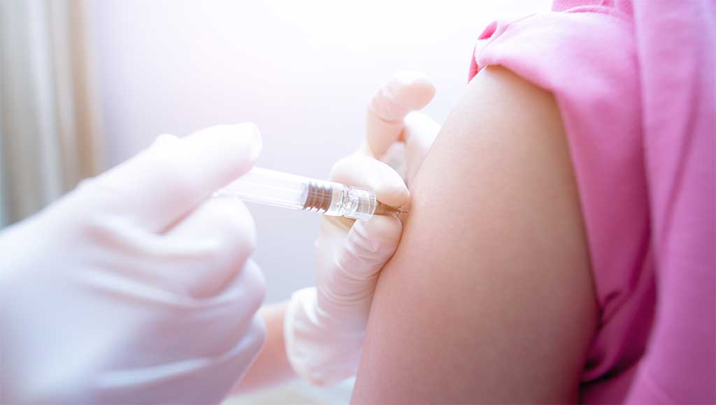
- Phototherapy – solar or UV radiation. Used in severe psoriasis; and
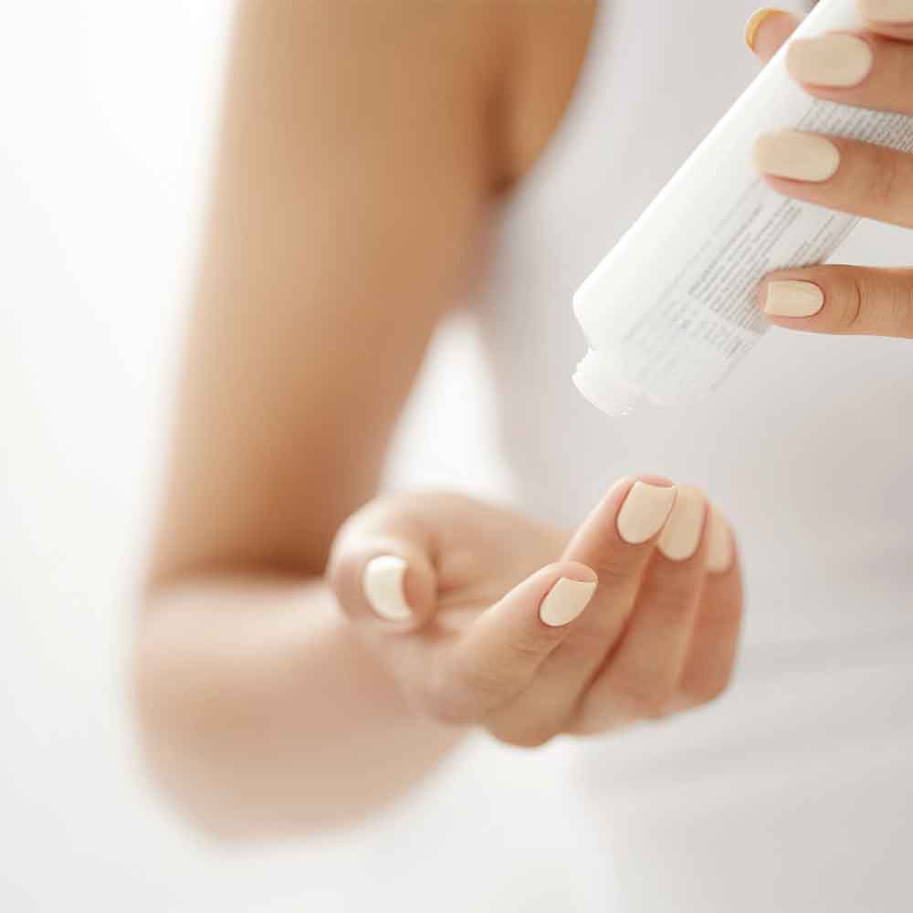
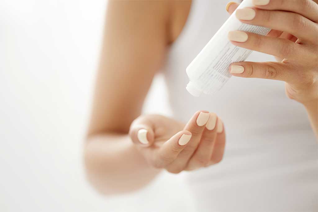
- Skin hydration by using emollients to keep the psoriatic skin soft and moist, hence reducing itch and tenderness.
These measures can be used alone in mild psoriasis or in conjunction with pharmacotherapy in moderate to severe cases.
Mild psoriasis may be treated with topical agents. Systemic treatment and phototherapy may be required in moderate to severe psoriasis.
Choice of topical treatment depends on location, characteristics of the lesions and patient preference.
Topical Corticosteroids are most widely used due to their known efficacy, ease of use and rapid onset of action. Occlusive dressings can be applied over small areas for a short period to increase the effectiveness of the topical steroid. One FTU (fingertip unit) is enough to cover an area twice the size of an adult hand. Use for the shortest possible time. Systemic absorption can occur, and, hence steroid strength needs to be monitored. Abrupt withdrawal of potent topical corticosteroids can cause rebound flare-up. Dose tapering is recommended prior to withdrawal.
Coal tar cream, gel, ointment, or foam can be applied 1-4 times a day.
Coal tar shampoo is used once a week. Massage into scalp, leave for several minutes and rinse.
Patient compliance may be a problem with coal tar products due to its odour, staining and sensitising properties. Coal tar can have a synergistic action with topical corticosteroids or phototherapy. Salicylic acid is often used in combination with coal tar as it acts as a keratolytic to soften and reduce psoriatic scale.
Salicylic acid cream or ointment can be applied 2-3 times a day.
Salicylic acid lotion is massaged into the scalp and then rinsed after 15 minutes. This is often used twice a day.
Salicylic acid shampoo can be applied twice a week to wet hair and rinsed after 3-5 minutes.
Calcipotriol 0.005% is available as a combination with betamethasone dipropionate 0.05% (Daivobet®). There is a theoretical risk of hypercalcaemia if used over large areas for long periods due to its action on vitamin D. A maximum of 100g of product per week is recommended. It is recommended that salicylic acid treatment be applied at a different time of the day to calcipotriol. Calcipotriol is unstable when used with salicylic acid.
Dithranol ointment or paste 0.05-2% is only available as an extemporaneous product. Start with a low strength for 5-10 minutes daily. Increase strength and application time (up to 30 minutes for short-contact treatment) according to the response.
Dithranol products are often combined with salicylic acid to improve stability. Combining it with coal tar helps reduce the irritant effect of dithranol.
Dithranol products are not used on sensitive areas like the face due to its irritant property, and it is applied directly to the lesion using gloves.
A calcineurin inhibitor (e.g. pimecrolimus) is effective for application to sensitive areas. It is less effective than topical corticosteroids.
Retinoids (tazarotene) are rarely used. Irritation is common.