Pityriasis lichenoides is a rare skin disorder of unknown cause, which can present in both an acute form (pityriasis lichenoides et varioliformis acute (PLEVA)) and a chronic form (pityriasis lichenoides chronica (PLC)) with many patients showing overlapping features of both (1,2). PLEVA can also evolve into PLC (1).
Although the cause is unknown, it is hypothesised to occur in the setting of a recent bacterial, viral or parasitic infection (2), or an inflammatory reaction to some medications, such as vaccines, or that it may be a low-grade lymphoproliferative disorder (1).
The skin disease most commonly tends to affect children and young adults under 30 years of age, with a slight male predominance. However, all ages and races can be affected, and it does not appear to be hereditary (1,2).
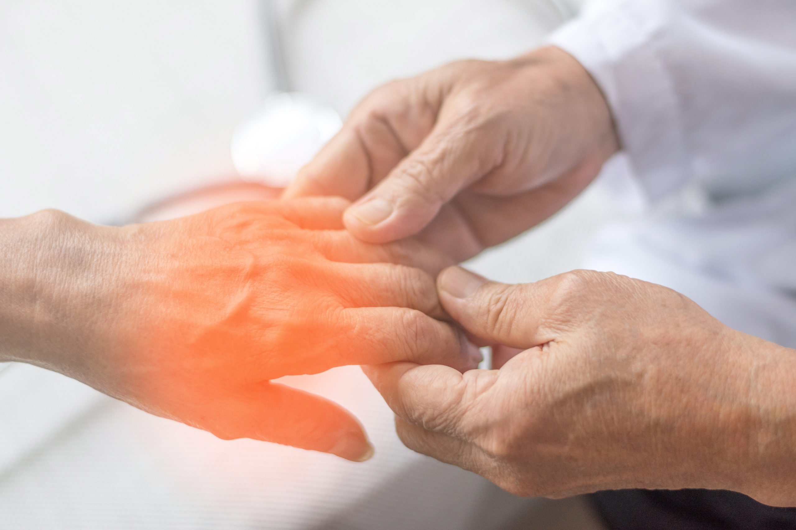
Pityriasis lichenoides can be difficult to diagnose, and diagnosis is often made on clinical grounds but is usually also confirmed with a skin biopsy (1,2). Referral to a dermatologist is important.
The clinical features and histological features of PLEVA and PLC are summarised below:
| PLEVA | PLC |
|
Clinical features |
|
| Rapidly progressive rash, but typically resolves within a few weeks, or can evolve into PLC | Presents more slowly over several days, but can last several months and wax and wane for several years |
| 10-50 pinkish or reddish or brownish flat spots, around 5-15mm in diameter | The spots look less red or inflamed than in PLEVA but are covered with a firm shiny scale of skin |
| Rash presents mostly on trunk, arms and legs, but in children they may appear more on the face than on other areas of the body | The scale can be scraped off by a dermatologist to reveal a shiny, reddish brown or discoloured surface underneath |
| Rash evolves into vesicles, pustules, hemorrhagic crusts and ulcers and most lesions heal with transient or persistent hyper or hypo pigmentation | The spots usually flatten within 3-4 weeks and the scale becomes loose, often leaving marks which appear darker than the person’s skin colour but these marks gradually fade away |
| Spots can come up at different times so the rash often consists of spots at various stages of development | Lesions can appear at various stages of evolution |
| Itchiness or burning sensations can be present | |
|
Histological features |
|
| A wedge-shaped deep dermal and superficial lymphohistiocytic infiltrate | A superficial dermal infiltrate |
| Parakeratotic scale and crust, with thinning of the granular layer | Focal parakeratosis |
| Interface dermatitis with basal cell necrosis and vacuolation | Preservation of the granular layer |
| Epidermal spongiosis and necrosis in more developed lesions | Focal loss of the dermo-epidermal interface |
| Extravasated erythrocytes | |
Pityriasis lichenoides may present as a rare form known as Febrile Ulceronecrotic Mucha-Habermann Disease, which has systemic features of malaise, fever, lymphadenopathy, arthritis and/or bacteraemia. There may also be mucosal, gastrointestinal, and pulmonary involvement and mortality of up to 25% has been reported (1). Psychological implications should also be considered, as skin lesions may appear on more visible areas such as the face (1).
There are no randomised controlled trials for the treatment of pityriasis lichenoides. However, various treatments exist, with varying levels of efficacy (1,2):
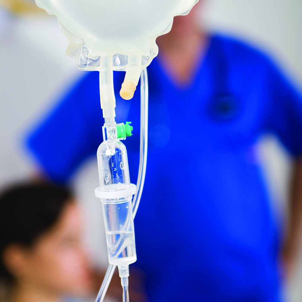
- PLEVA is most commonly treated with a three-month course of erythromycin in younger children or with doxycycline. These are used primarily for their anti-inflammatory effects, rather than their antibacterial properties. Erythromycin has been used at a dose of 30 to 50mg/kg per day given in three to four divided doses for one to four months, however a dermatologist should guide treatment doses (3).
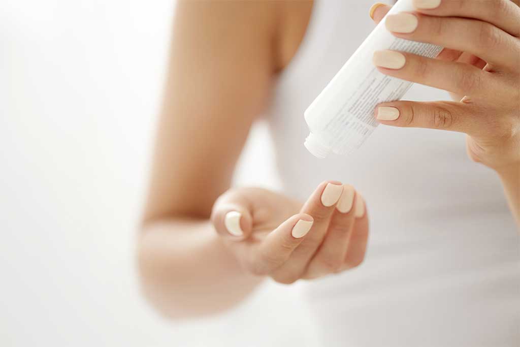
- Topical corticosteroids and topical tacrolimus may relieve symptoms but do not make the rash disappear more quickly
- Phototherapy with ultraviolet-B is often the preferred treatment for PLC.
- Methotrexate and other immunosuppressants may be considered for refractory or very severe cases.
- Antihistamines may be used to reduce itching.